Research
Overview
The lab develops computational methods that combine quantitative MRI, inverse problems, machine learning, and clinically motivated neuroimaging analysis.
Current Directions
Accelerated MRI
Fast acquisition and reconstruction methods for quantitative imaging, with an emphasis on practical deployment, robustness, and translational impact.
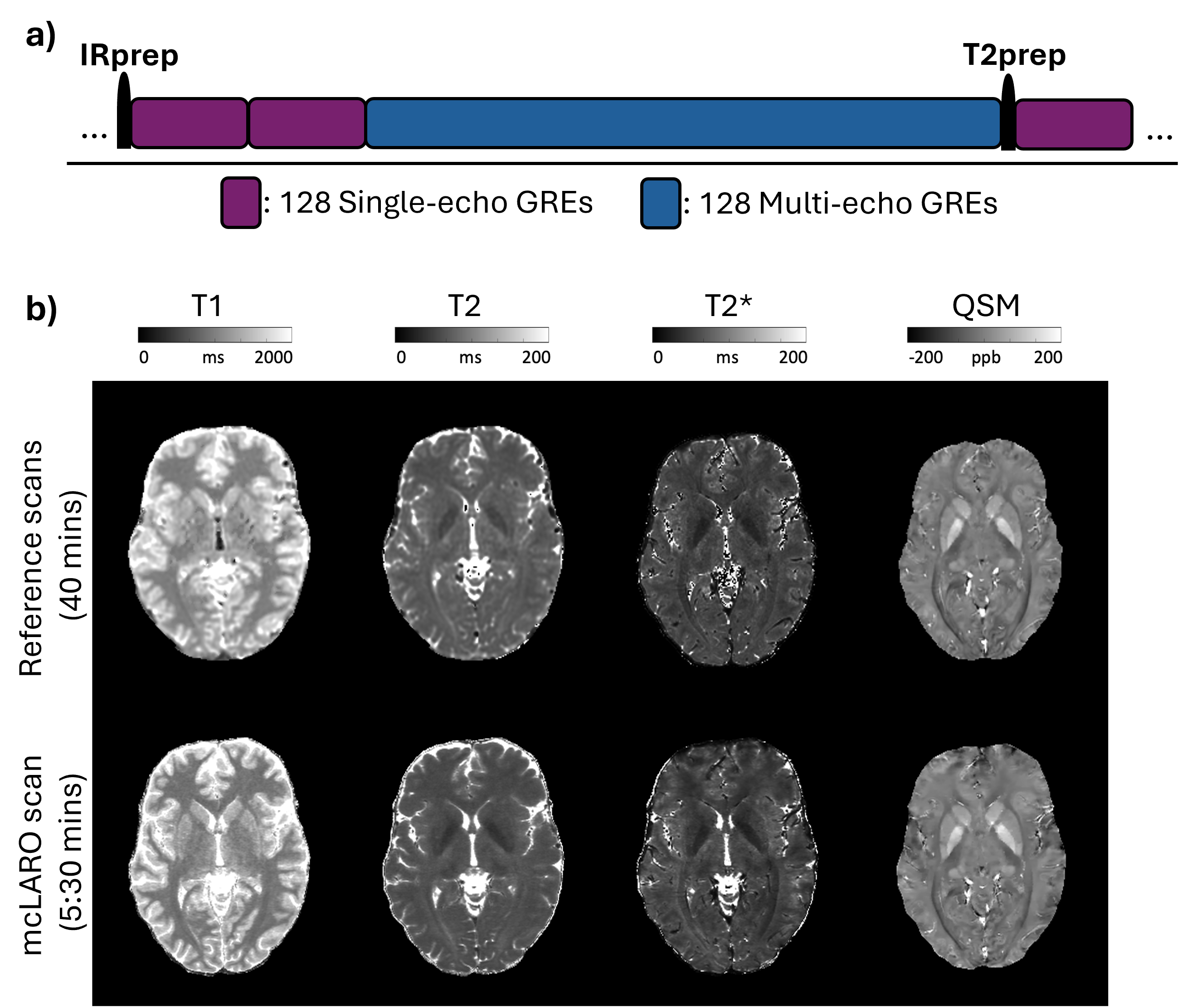
(a) mcLARO pulse sequence diagram. (b) Comparison of mcLARO qMRI maps with reference standards.
Related Publications
- LARO: Learned acquisition and reconstruction optimization to accelerate quantitative susceptibility mapping. NeuroImage, 2023.
- mcLARO: Multi-contrast learned acquisition and reconstruction optimization for simultaneous quantitative multi-parametric mapping. Magnetic Resonance in Medicine, 2024.
- Navigator motion-resolved MR fingerprinting using implicit neural representation (FINR): feasibility for free-breathing 3D whole-liver multiparametric mapping. Magnetic Resonance in Medicine, 2025.
- Spiral cardiac quantitative susceptibility mapping for differential cardiac chamber oxygenation: initial validation in relation to invasive blood sampling. Magnetic Resonance in Medicine, 2025.
Quantitative Tissue Parameter Mapping
Methods for quantitative tissue parameter estimation, reconstruction, and downstream analysis in neurological applications, especially where quantitative tissue characterization matters.
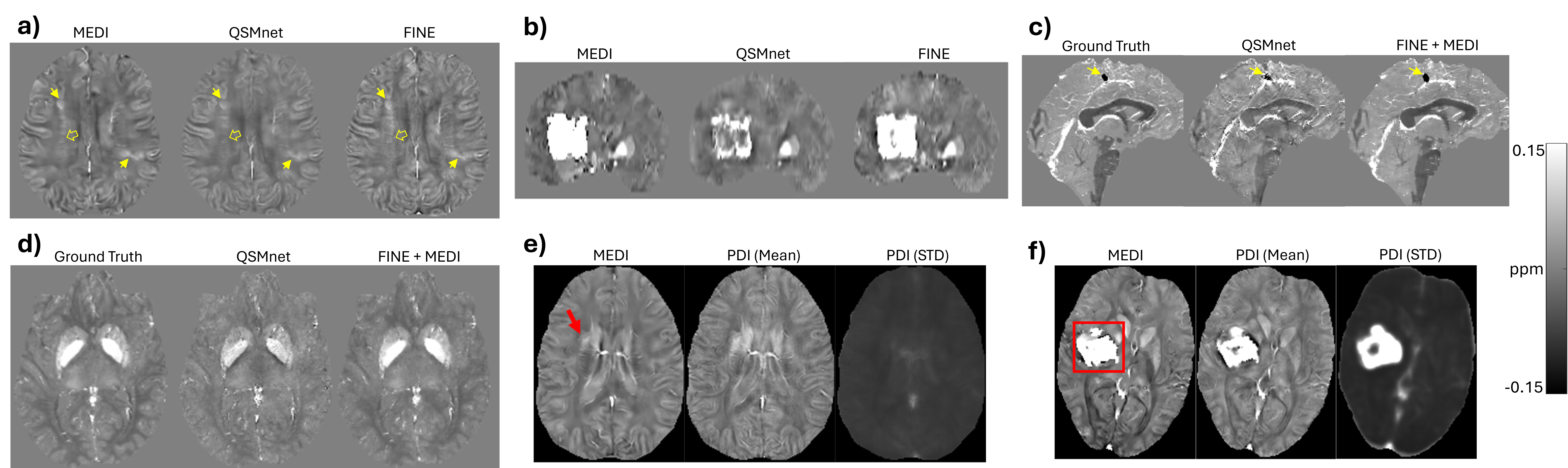
QSM reconstructions for representative cases: (a) an MS patient and (b) an ICH patient using MEDI, QSMnet, and FINE; (c) a calcification case and (d) a low-SNR case from the 2020 QSM Challenge 2.0 using QSMnet and FINE+MEDI; and (e) PDI reconstructions of an MS and an ICH patient, showing susceptibility mean and standard deviation (STD) maps.
Related Publications
- Fidelity imposed network edit (FINE) for solving ill-posed image reconstruction. NeuroImage, 2020.
- Probabilistic Dipole Inversion for Adaptive Quantitative Susceptibility Mapping. Machine Learning for Biomedical Imaging, 2021.
- QQ-NET: using deep learning to solve quantitative susceptibility mapping and quantitative blood oxygen level dependent magnitude based oxygen extraction fraction mapping. Magnetic Resonance in Medicine, 2022.
- Deep neural network for water/fat separation: supervised training, unsupervised training, and no training. Magnetic Resonance in Medicine, 2021.
Lesion Analysis
Automated segmentation, longitudinal tracking, and biomarker discovery for white matter lesion burden and disease progression.
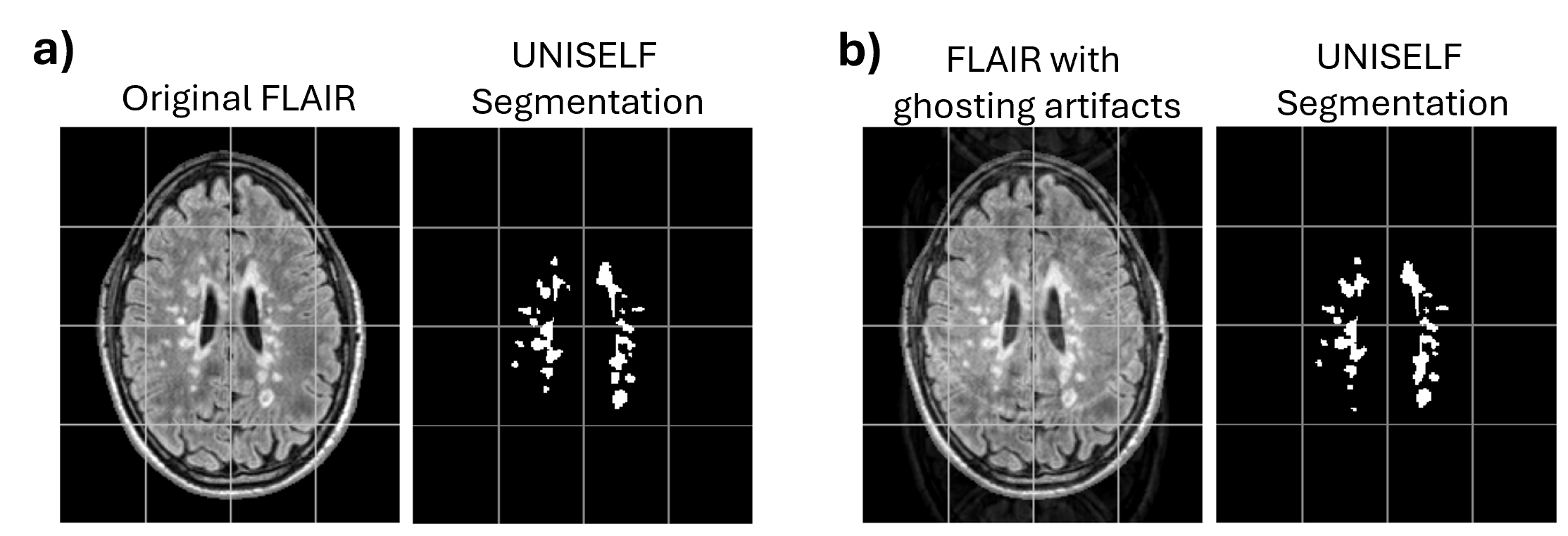
UNISELF segmentations on FLAIR: (a) original vs. (b) with ghosting artifacts.
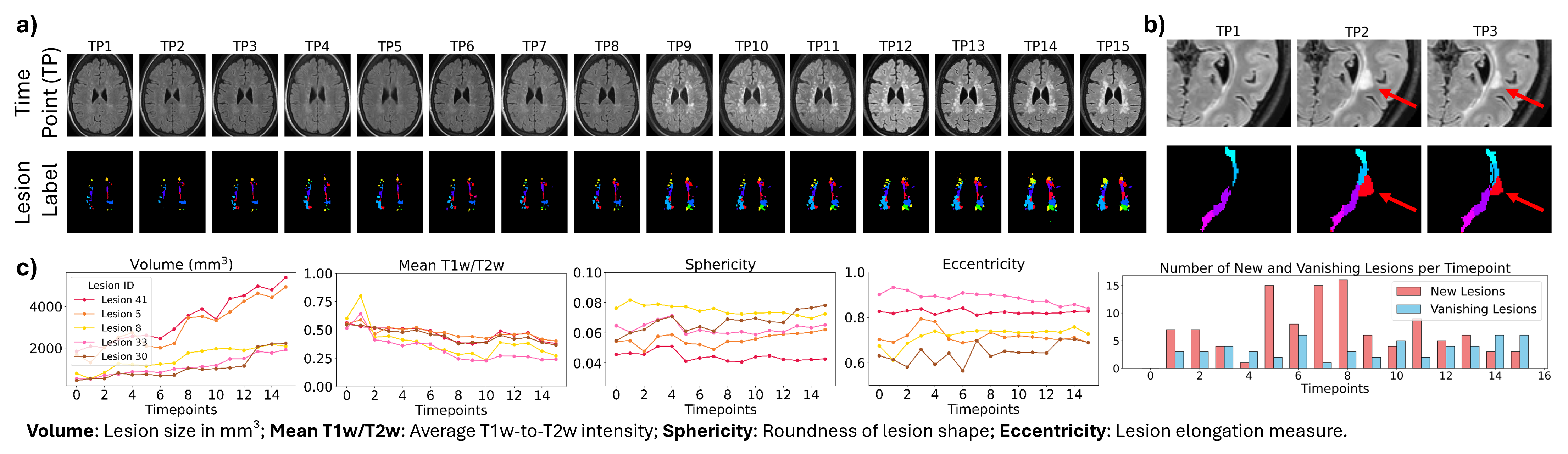
(a) Example of unique MS lesion labels across fifteen timepoints extracted by AULTRA. (b) Zoom-in view showing the separation and tracking of a new confluent lesion (red arrows) across three timepoints. (c) Unique lesion characterization over time, visualizing longitudinal changes (x-axis) in the five largest enlarging lesions from (a).
Related Publications
- UNISELF: A Unified Network with Instance normalization and Self-Ensembled Lesion Fusion for Multiple Sclerosis Lesion Segmentation. Medical Image Analysis, 2026.
- Bi-directional MS lesion filling and synthesis using denoising diffusion implicit model-based lesion repainting. SPIE Medical Imaging, 2025.
- ALL-Net: Anatomical information lesion-wise loss function integrated into neural network for multiple sclerosis lesion segmentation. NeuroImage: Clinical, 2021.
- QSMRim-Net: Imbalance-aware learning for identification of chronic active multiple sclerosis lesions on quantitative susceptibility maps. NeuroImage: Clinical, 2022.